
Understanding Your Breath: Why These Numbers Matter
You've been coughing for weeks, and your doctor finally ordered those papers and tubes you breathe into. Now you have a report full of acronyms like FEV1, FVC, and DLCO staring back at you. What does it actually mean? You aren't alone in feeling confused by these charts.
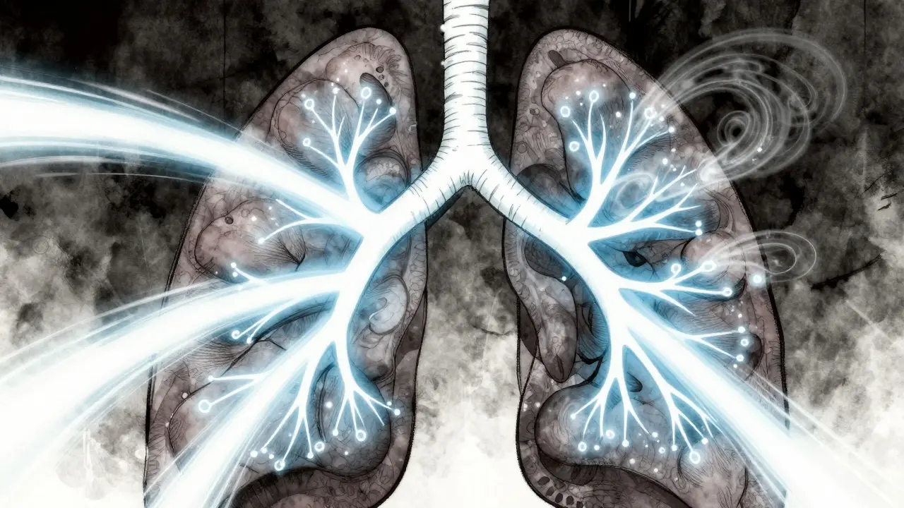
Pulmonary function testing tells us exactly what your lungs are doing when they take a deep breath and blow out hard. It is often the most reliable way to figure out if your shortness of breath comes from narrowed airways, stiff lungs, or something else entirely. While many patients assume these tests are just about "airflow," the reality is much deeper. We need to look at volume, speed, and how well your blood absorbs oxygen across that delicate membrane inside your lungs.
In this guide, we break down the jargon into plain English so you can walk into your next appointment with questions that matter. We will explore how Pulmonary Function Tests serve as a window into your respiratory health, offering concrete data on capacity and efficiency.
The Basics of Spirometry: Flow and Volume
Spirometry is the most common part of the exam, and honestly, it is the most misunderstood. Most people know the test involves blowing into a machine, but few understand the two main numbers being recorded: Forced Vital Capacity (FVC) represents the total amount of air you can blow out after taking the deepest breath possible. Think of FVC as the size of your tank. A healthy adult might have a capacity of around 4 to 5 liters, depending on their height and age.
The second number, Forced Expiratory Volume in 1 Second (FEV1), is the volume of air you can blast out in the very first second of that exhale. This measures the speed of your airflow. Imagine a fire hose; if the water comes out fast, the hose is open. If it dribbles out slowly, there is a blockage. FEV1 checks that blockage. The magic lies in the ratio between the two, written as FEV1/FVC.
Clinical Rule of Thumb:
- If the FEV1/FVC ratio drops below 0.70, it suggests Obstructive Lung Disease, such as asthma or COPD, where the pipes get narrow.
- If the FEV1/FVC ratio stays above 0.70 but the total volume (FVC) is low, it points toward restriction, meaning your lungs cannot fully inflate.
- Normal values generally range between 80% and 120% of what is predicted for someone your size.
Decoding DLCO: The Gas Exchange Test
This is where things get interesting. Spirometry tells us about the pipes, but DLCO tells us about the engine's fuel intake. Diffusing Capacity of the Lungs for Carbon Monoxide (DLCO) is a test that measures how effectively oxygen moves from your air sacs (alveoli) into your bloodstream. We use carbon monoxide for the test because it binds to hemoglobin so strongly that we can track exactly how much crosses over the barrier.
Unlike spirometry, which you can guess roughly by effort, DLCO is purely physiological. The technician asks you to hold your breath for ten seconds after inhaling a specific gas mix. During that pause, the carbon monoxide travels across the alveolar-capillary membrane. We calculate the result in milliliters per minute per mm Hg, adjusted for your hemoglobin levels.
| Pattern | FVC Result | FEV1/FVC Ratio | DLCO Result | Common Causes |
|---|---|---|---|---|
| Obstruction | Normal or Low | < 0.70 (Low) | Variable (Often Low in Emphysema) | Asthma, Chronic Bronchitis, COPD |
| Restriction | Low (< 80%) | Normal (> 0.70) | Reduced | Interstitial Lung Disease, Sarcoidosis |
| Normal Restriction | Low | Normal | Normal | Obesity, Chest Wall Abnormalities |
| Vascular Issue | Normal | Normal | Low | Pulmonary Hypertension, Embolism |
This distinction is critical. You could have severe emphysema where spirometry looks okay, but DLCO reveals the damage immediately. Conversely, if you are obese, spirometry might show low volumes (restriction), but DLCO remains high because the actual lung tissue is fine; your chest just can't expand fully.
Interpreting Complex Patterns
Lung disease is rarely simple, and sometimes the tests overlap. Here is how expert pulmonologists differentiate the tricky cases using the combined data from these lung function tests.
Consider the scenario where spirometry suggests obstruction but DLCO is perfectly preserved. This is classic Asthma or chronic bronchitis. The airways are inflamed or mucus-filled, narrowing the tube, but the surface area for gas exchange remains intact. The damage hasn't eaten away the alveoli yet.
Now, look at the opposite: Spirometry shows restriction (low FVC), and DLCO is also significantly reduced. This combination almost always signals interstitial lung disease or fibrosis. The scar tissue in the walls of the lungs makes them stiff (low volume) and thickens the membrane, making oxygen transfer inefficient. We often see the DLCO drop before spirometry picks up the change, serving as an early warning sign months before you feel breathless during exercise.
There is a third, less obvious pattern: Normal spirometry but low DLCO. This is the hallmark of vascular issues. If your airways are open and your lung capacity is fine, why isn't oxygen getting in? It usually points to Pulmonary Hypertension or chronic emboli. Blood flow is blocked, not air flow. This specific finding saves lives because treating high blood pressure in the lungs differs vastly from treating asthma.

Factors That Skew Your Results
Even the best test can give a misleading picture if external factors aren't accounted for. The American Thoracic Society guidelines emphasize correcting for several variables to ensure accuracy.
First is hemoglobin. Anemia lowers DLCO simply because there are fewer red blood cells to carry the gas, mimicking lung disease. For every gram per deciliter of hemoglobin below normal, DLCO drops about 7%. If your lab work showed low iron the week before, your DLCO might look terrible even if your lungs are healthy.
Second, altitude plays a role. People living in cities like Denver or Mexico City naturally have lower DLCO readings due to lower atmospheric pressure. Reference ranges adjust for this, but a local clinic running standard sea-level equations might flag a mountain resident as having "abnormal" results incorrectly.
Finally, smoking has a unique effect. Smokers accumulate carboxyhemoglobin, which blocks hemoglobin sites. Paradoxically, heavy smokers sometimes show a higher DLCO than non-smokers because the body produces more red blood cells to compensate for hypoxia, while the carbon monoxide binding creates a false high reading initially, though long-term damage eventually crushes the numbers. Ideally, you should stop smoking at least three hours before the test to get a true baseline.
Your Questions Answered
What does a low FEV1/FVC ratio really mean?
A low ratio means your airways are obstructed. It indicates that air is trapped behind a bottleneck and cannot exit quickly enough. This is the defining feature of COPD and asthma.
Can I do a DLCO test if I have just flown recently?
No, avoid the test within 3 days of air travel or scuba diving. High-altitude flights increase red blood cell counts slightly, which artificially elevates DLCO values.
Is spirometry painful or dangerous?
It is non-invasive and painless, though the forceful breathing can trigger mild coughing or dizziness. It is safe for almost everyone unless you have had recent eye or abdominal surgery.
Why is my FVC low but my DLCO is high?
This usually indicates extrinsic restriction, like obesity or muscle weakness. Your lung tissue itself is healthy (high DLCO), but your rib cage cannot expand fully to pull air in (low FVC).
How often should I repeat these tests?
Frequency depends on the condition. Stable COPD is checked annually. Progressive fibrosis might require checks every 3-6 months to monitor decline rate against therapy.